- About Us
Fact SheetOverseas Offices & RepresentativesAccreditations and AwardsPatient Stories
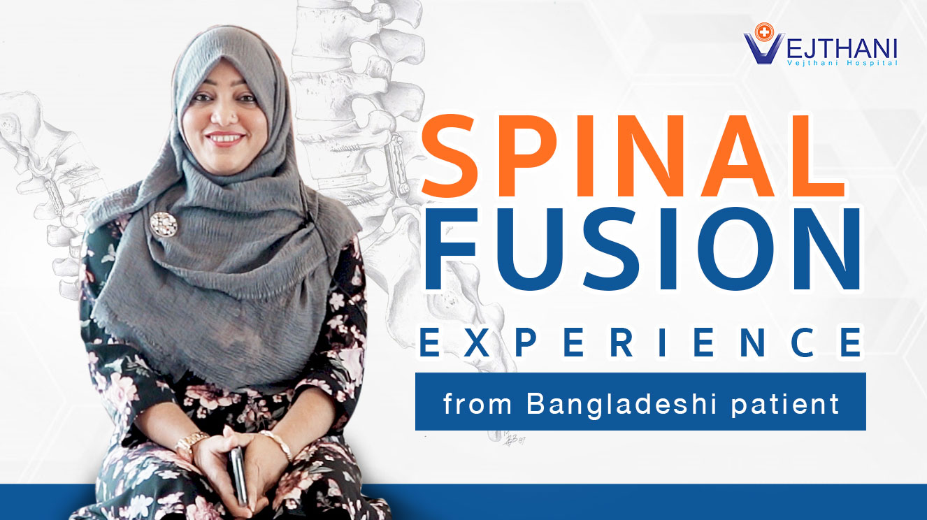
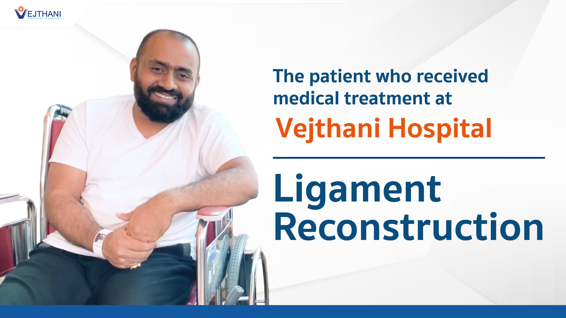
 Spinal fusion experience from Bangladeshi patientMyanmar Actress Patricia’s Intraocular Lens Surgery Experience at Vejthani HospitalLigament ReconstructionA comprehensive kid’s checkup at Vejthani Hospital’s Super Kid’s Center
 - Patient Services
- Medical Departments & Centers
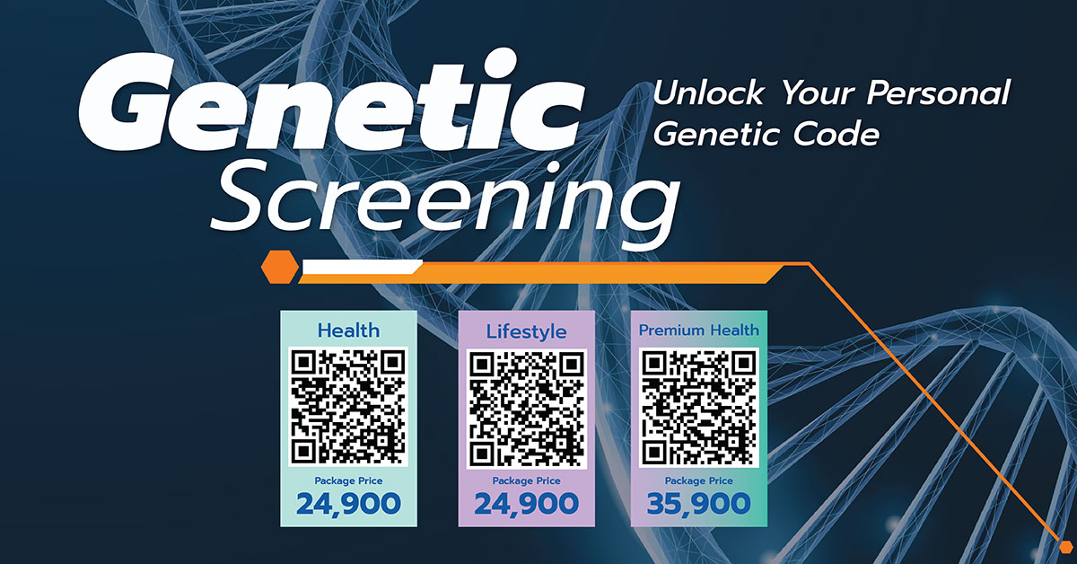
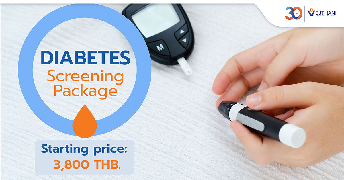
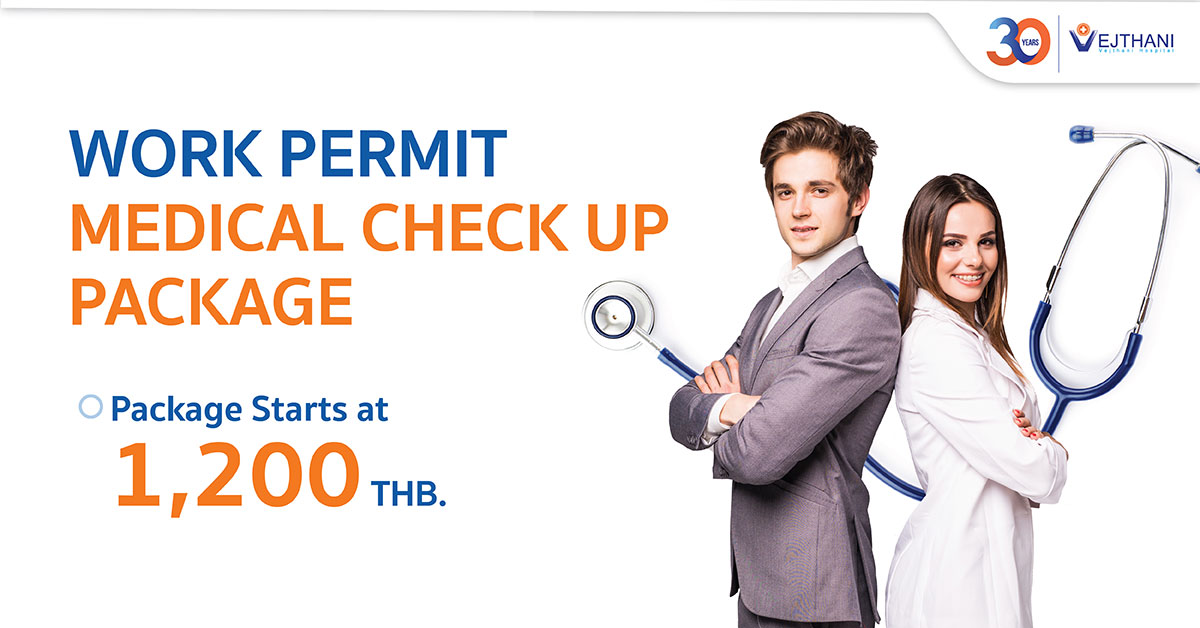
- Packages & Programs
- Health Information
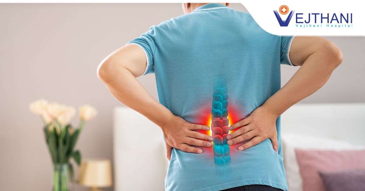
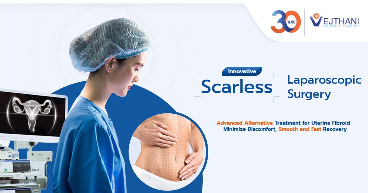
Health ArticlesHealth VideosNews and UpdatesTreating Chronic Back Pain that Could Be a Sign of Herniated DiscSummer’s Silent Killer: Your Essential Guide to Preventing and Defeating HeatstrokeGroundbreaking Approach to Treat Cerebral Aneurysm: Biplane DSA and the Future of NeurointerventionScarless Laparoscopic Surgery: Advanced Treatment for Uterine Fibroid


- About Us
Fact SheetOverseas Offices & RepresentativesAccreditations and AwardsPatient StoriesSpinal fusion experience from Bangladeshi patientMyanmar Actress Patricia’s Intraocular Lens Surgery Experience at Vejthani HospitalLigament ReconstructionA comprehensive kid’s checkup at Vejthani Hospital’s Super Kid’s Center
- Patient Services
- Medical Departments & Centers
- Packages & Programs
- Health Information
Health ArticlesHealth VideosNews and UpdatesTreating Chronic Back Pain that Could Be a Sign of Herniated DiscSummer’s Silent Killer: Your Essential Guide to Preventing and Defeating HeatstrokeGroundbreaking Approach to Treat Cerebral Aneurysm: Biplane DSA and the Future of NeurointerventionScarless Laparoscopic Surgery: Advanced Treatment for Uterine Fibroid
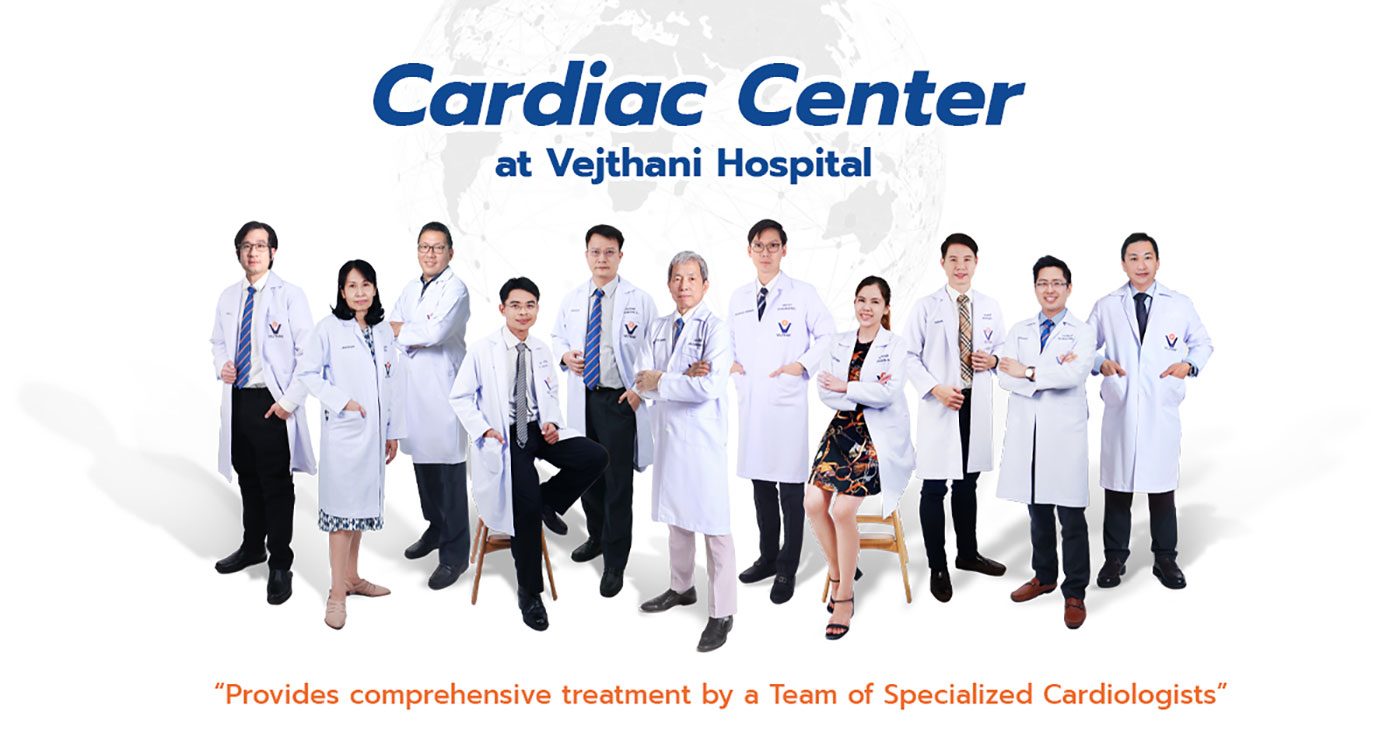
Cardiac Center
CARDIAC CENTER
Cardiac Center at Vejthani Hospital in Bangkok is more than willing to serve you with our finest heart checkups and cardiac treatment. As we understand patients with a cardiac disease do not have an easy or pain-free experience, our Heart Center in Bangkok, Thailand, carefully selects only the latest advanced technologies and facilities to support operations and ease the pain of our valued patients.
Our team of surgeons consists of thoroughly experienced, dedicated and certified cardiac specialists so you can rest assured that you will receive professional cure by our qualified and caring professionals. The service is of a world-class standard where the staff are well-trained and truly care for your health and needs. You are warmly welcomed to Vejthani Cardiac Center in Bangkok, Thailand, where our team of cardiac experts with friendly customer service and medical staff are more than ready to serve you.
SERVICE HOURS
Monday – Sunday : 07.00 am – 08.00 pm
LOCATION
Cardiac Center, 5th Floor, Vejthani Hospital
APPOINTMENTS & INQUIRIES
Call : +66 (0) 2734-0000 ext. 5300
Fax : 02-734-0000 ext. 5323
Services
1) Non-invasive investigations include
- Electrocardiography (ECG or EKG)
- Ankle-brachial Index
- Exercise Stress Test
- Echocardiogram
- Holter Monitoring
- Ambulatory Blood Pressure Monitoring
- Dobutamine Stress Echocardiography
- Transesophageal Echocardiogram
- Exercise Stress Echocardiography
- Assessment of Syncope
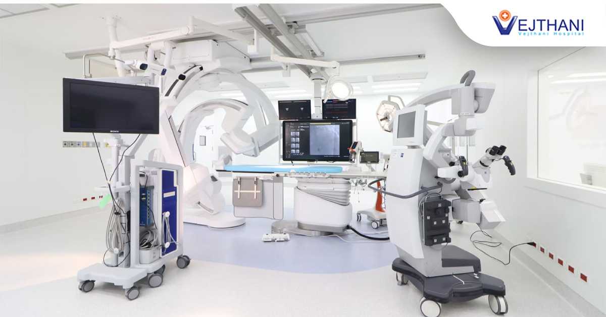
2) Cardiac cath lab
- Peripheral Angiography
- Percutaneous Coronary Interventions and Stents
- Permanent Pacemaker Implantation
- Electrophysiologic Study and Radiofrequency Ablation
- Atrial Septal Defect Device Closure
- Percutaneous Mitral Balloon Valvuloplasty
- Automated Implantable Cardiac Defibrillators (AICD)
- Intra-aortic Balloon Pump Insertion (IABP)
3) Cardiothoracic and Vascular Surgery
- Coronary Artery Bypass Graft
- Valvular Repair / Replacement Surgery
- Congenital Heart Defect Surgery
4) Cardiac Ward specifically for cardiac patients with monitoring equipment to monitor the rates and rhythms of heartbeats, the circulatory system and patient conditions around the clock
5) Coronary Care Unit equipped with necessary equipment to closely monitor critical cardiac patients under the universal standard
6) Cardiac Rehabilitation which provides consultations and knowledge regarding heart diseases and behavioral adjustment, together with personalized workout plans to support heart function recovery as well as nutritional advice for heart disease patients
7) Cardiac Computed Tomography
8) Cardiovascular Magnetic Resonance Imaging: (CMR)
9) Mobile Coronary Care Unit for critical cardiac patients